Magnetic Resonance Imaging
Jim Kelly
Contents
·
Introduction
·
What is NMR ?
·
A Medical Marvel
·
Vocabulary
·
The Technique I & II
·
Magnet Parameters
·
The Applications
·
Summary
· References
Introduction
·
NMR Imaging was
developed by Lauterbur & Mansfield in the early 1970’s
·
Initial poor image
quality meant No Medical Relevance, with development seen as a
New Medical Revolution
What is NMR ?
·
A technique developed
over 40 years ago as an analytical chemical tool
·
It works because some
nuclei are weakly magnetic and respond to magnetic fields
·
The nuclei are then
excited by an applied radio frequency ( R.F. ) pulse and radiate energy
·
This yields information
about the physical and chemical composition of the sample
·
Varying the magnetic
field gives spatial NMR images of slices of the object
· Most non metallic materials are transparent to to magnetic fields and radio frequencies
A Medical Marvel
· Non invasive imaging capability sensitive to
subtle differences in water content detects local flow characteristics of onset
of for example: multiple sclerosis, cancer, cardio-vascular conditions etc.
·
routine brain scans
possible
·
novel applications with
arthritis
· whole body imaging with non ionising radiation possible
Vocabulary
·
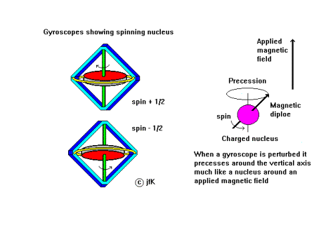
Gyro; To rotate, a gyroscope can precess
·
Gyromagnetic ratio; G = 42.571 MHz T-1 for H
·
Larmor frequency; f = (G/ 2 p) B, the rate of precession of nuclei in
a magnetic field
·
Precession; To rotate the axis of a spinning body at a angle
about a given direction
·
Relaxation time; The time T1 for spins to re-align
themselves in the direction of the field, for
T2 the spin-spin interaction to decay
·
Spin; The angular momentum e.g. of a proton
·
Tesla; unit of magnetic field strength B
The Technique I
·
The hydrogen nucleus has
a charge and angular spin so it acts like a tiny bar magnet
·
Nuclei in water will
therefore align themselves in an external magnetic field
·
A radio frquency pulse
at the Larmor frequency f L will tilt the nuclei from this direction
·
They will then precess about the
direction of the field losing energy as radio waves
·
Coils around the sample
detect an induced voltage as the strength of the signal decays
· The T1 relaxation time for a compass needle to come to rest
The Technique II
·
The T2
relaxation time (spin-spin interaction) for compass needles to come to rest
·
The spins exchange
energy resulting in a longer relaxation time than the spin-lattice T1
·
Signals in the coil are
detected and analysed into frequency components
·
Spatial information
obtained using a magnetic field gradient, built up in 2D slices
·
This gives a map of
water content in tissue, bone appears dark and soft tissue bright
·
Both T1 &
T2 are longer in diseased tissue
Magnet Parameters
·
Superconducting
electromagnet of Ni-Ti Alloy used
·
Uniform Field strengths
of between 2 to 5 Tesla, Field gradients stable to @ 1%
·
Machines cost about £1
Million
·
Whole body machines are
becoming smaller
The Applications
·
Imaging tumours in the
brain
·
Measurement of the fat
content of foods
· Determination of the chemical composition of substances
Summary
·
How does MRI work and
what use is it ?
·
Calculate the precession
frequency of hydrogen in a magnetic field of 2 Tesla
·
At what temperature are
the coils in the electromagnet maintained ?
· What is the difference between a sagital and transverse scan ?
References
·
“The Industrial
Applications of MRI” The Royal Society New Frontiers in Science 1996
·
“Medical Physics” by
Martin Hollins p186, published by Nelson
·
MRI resource pack
produced by the University of Surrey, Physics Dept.
· Chemistry in Britain vol 32 no 6 p33-45 June 1996
