 |
Laboratory X-ray Sources |
|
|
Laboratory X-ray Sources |
Laboratory X-ray Sources
Of the two experimental methods for creating X-rays in the laboratory, only the sealed-glass X-ray tube is in common use for powder diffraction studies. In recent years, the sealed glass tube has been improved by the use of ceramics, but otherwise the two types are very similar. The latest developments have been the production of microfocus tubes, but their use is still not established in the powder diffraction community, probably because of cost and as yet unproven reliability.
Sealed Glass X-ray Tube
Shown below is a picture of a typical sealed glass X-ray tube. You may like to compare this to the museum photograph.
 |
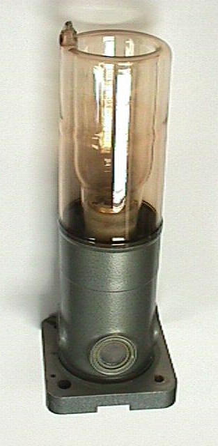 |
The windows are made of beryllium metal which has a very low absorption cross-section for X-rays. The tube shown here has a copper anode and electrons are produced by heating a tungsten filament. If you wonder what the inside of an X-ray tube actually looks like, then take a look at the photographs.
The schematic diagram below emphasises the key aspects behind the operation of the X-ray tube. The exact operating voltage of the tube depends on the characteristics of the filament, but generally the heat load on the anode is of the order of kilowatts. Most of the energy of the electrons hitting the anode target is dissipated as heat and so the tube is water cooled as indicated in the diagrams. X-rays are generated in all directions, but the optimum angle for viewing the source is typically at about 6° to the anode surface.
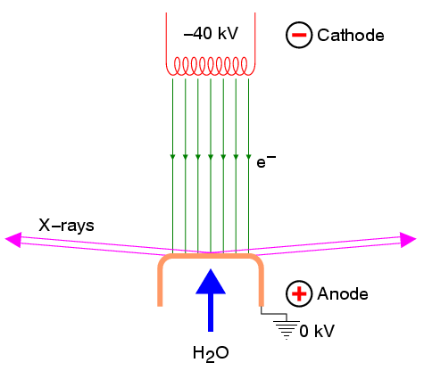
The final point to make is that you may have noticed that the X-ray tube has four windows. Two of these are parallel to the filament and the other two are perpendicular to it. If a spot source of X-rays is required, then the tube should be arranged so that a window parallel to the filament axis is used, in contrast to when a line source of X-rays is required when a window perpendicular to the filament axis is used. Note that most modern powder diffractometers make use of a line source in contrast to the older Debye-Scherrer cameras which made use of the spot source.
| © Copyright 1997-2006. Birkbeck College, University of London. | Author(s): Jeremy Karl Cockcroft |